
Use of 3D reconstruction cloacagrams and 3D printing in cloacal malformations
Jennifer Ahn, MD, Margarett Shnorhavorian, MD, MPH, Giri Shivaram, MD, Ray Ramoso, RT, Anne-Marie Amies Oelschlager, MD, Jeffrey Avansino, MD, Paul Merguerian, MD, MS.
Seattle Children's Hospital, Seattle, WA, USA.
Introduction: Cloacal anomalies are rare and require complex inter-disciplinary care. Anatomic findings such as length of common channel, Mullerian structure development, and bladder size are essential in operative planning and family counseling regarding long-term bowel, bladder, and sexual function. We present our experience with the use of 3-dimensional (3D) reconstruction cloacagrams and 3D printing for patients with cloacal and urogenital sinus anomalies.
Methods: A retrospective review of all patients undergoing 3D cloacagram for cloacal and urogenital sinus anomalies was performed. Each patient also underwent cystoscopy, vaginoscopy, and exam under anesthesia. Intra-operative measurements and findings were compared to measurements from the cloacagrams.
3D Cloacagram protocol: 6Fr or 8Fr foley catheters were placed into the mucus fistula, bladder, and vagina. Catheter balloons were inflated and lead BB markers were placed at the anticipated location of the anus and at the cloacal orifice. Catheters were instilled with iothalamate meglumine (Cysto-Conray II) under intermittent fluoroscopic observation to obtain maximal distention. The catheters were then clamped, and cone beam CT imaging (XperCT) of the pelvis was performed. Images were reconstructed to produce multiplanar and volume-rendered reformations. Measurements were performed of the common channel length, common channel to bladder, and distal colon to anus. Contrast was aspirated and catheters removed.
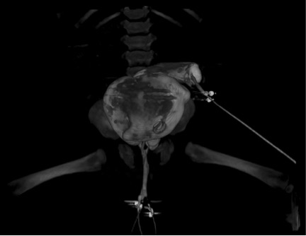
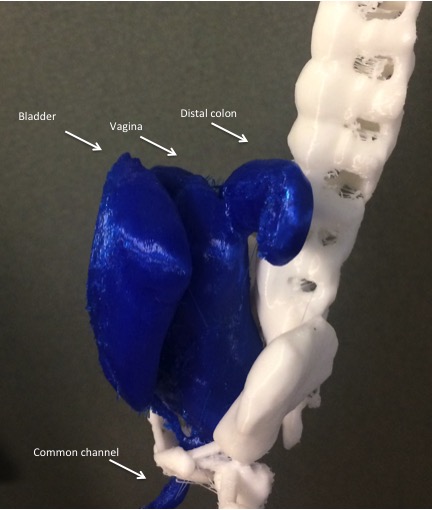
Results: Five patients underwent both endoscopy and 3D cloacagram, with four patients having both procedures done under the same anesthetic. Patient characteristics, intraoperative measurements, and cloacagram findings are shown in Table 1. Representative cloacagram images are seen in Figures 1-2. Figure 3 illustrates a 3D model printed from patient 3’s cloacagram; the 3D model was used for family education and surgical planning.
Conclusion: 3D cloacagrams provide measurements similar to endoscopic findings, and anatomic details which are helpful in preoperative planning. It also provides a platform for 3D printed models, which can be used for surgical simulation, as well as patient and trainee education.
Table 1 Patient characteristics and results of cloacagram and endoscopy
Figure 1 3D cloacagram
Figure 2 3D cloacagram with measurements
Figure 3 3D printed model of patient #3’s cloacagram 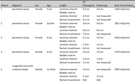

Back to 2016 Fall Congress