-->
|
Back to 2014 Fall Congress Meeting Abstracts
The double zipper theory of male anterior urethra development visualized in three dimensions by optical projection tomography
Yi Li, BA, Sisir Botta, MD, Adriane W. Sinclair, PhD, Gerald R. Cunha, PhD, Laurence S. Baskin, MD.
University of California, San Francisco, San Francisco, CA, USA.
Background
There are currently two schools of thought regarding the embryogenesis of the male urethra. Older literature most commonly refers to the ectodermal ingrowth theory proposed by Glenister, who described the concept of an invagination of ectodermal cells forming the stratified squamous epithelium of the glandular urethra. A contemporary theory of urethral development as described by Kuzrock is the endodermal differentiation theory, which argues that the glandular urethra is formed by growth of the urethral plate to the distal tip of the penis. Previous studies examining urethral development in human embryogenesis have relied primarily on serial section measurements and occasionally on 3D reconstruction of serial images. Herein we describe a novel approach to imaging human urethral development through whole mount staining of fetal genital tubercle with an E-cadherin antibody and immunofluorescent imaging with optical projection tomography (OPT). We hypothesize that OPT analysis will support the endodermal differentiation theory of urethral development.
Methods
Thirty one anonymous human fetal genital specimens were acquired after IRB approval. Gender was identified through PCR and gestational age estimated by heel-toe length. Specimens were fixed in formalin, processed for OPT, and stained with E-cadherin antibody. Stained specimens were sent to an imaging core for OPT imaging. The resulting data was then analyzed using Volocity software.
Results
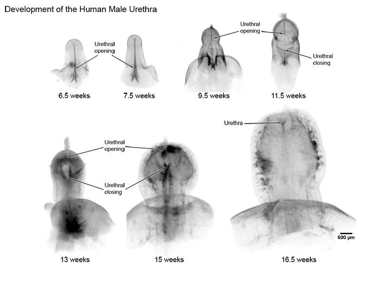
Seven developing male fetal specimens from 6.5 weeks gestation to 16.5 weeks gestation were processed for OPT and the remaining 5 male specimens analyzed by serial section. Length of phalluses ranged from 1.3mm to 3.7mm, respectively. We visualized the urethral opening moving proximal to distal as gestational age increased. In the 6.5 week specimen, the distal 2/3 of the urethra plate remained solid with evidence of proximal to distal canalization. In comparison, the 9.5 week specimen showed a urethral opening which extended distally to the coronal sulcus with no proximal closure. The 11.5 week specimen revealed an opening extending to the mid-glans and the urethra closing proximally to the mid-shaft. In the 13 week specimen, the urethral opening extended to the distal glans and had closed proximally to the coronal sulcus. Older samples showed continued distal extension of the urethral opening (Figure 1).
Conclusions
Herein we describe 3D visualization of a “double zipper”, wherein one zipper opens the urethral opening proximal to distal through canalization and unfolding of the urethral plate, and a second zipper follows behind and closes the urethral groove to form the urethral tube. This observation supports the endodermal differentiation theory with no evidence of ectodermal intrusion. We propose OPT as an improved technique for visualizing morphological development in fetal structures. Understanding normal urethral development is the first step in solving the mystery of hypospadias.
Back to 2014 Fall Congress Meeting Abstracts
|

