-->
|
Back to 2014 Fall Congress Meeting Abstracts
Histologic Properties of Autologous Buccal Mucosal Vaginoplasty
Gwen M. Grimsby, MD, Sandy Cope-Yokoyama, MD, Linda A. Baker, MD.
UT Southwestern Medical Center/Children's Medical Center, Dallas, TX, USA.
Introduction: Since buccal mucosa and native vagina are both moist stratified squamous epithelia, autologous buccal mucosa vaginoplasty (ABMV) has been recently used. However, important native vaginal properties, including presence of estrogen receptors (ERs), nerves, and Langerhans cells (LCs), the portal of entry of bacterial and viral infections, has not been studied. Defining these histologic characteristics may translate to improved patient counseling concerning risk of sexually transmitted infections (STIs), utility of hormonal therapies, and sexual sensation after buccal vaginoplasty. The goal of this study was to compare the innervation, presence of ERs, and density of LCs in ABMV buccal grafts versus the patient’s native vaginal tissues.
Materials and Methods: In this IRB approved prospective study from 2008 to present, ABMV patients underwent biopsy of their buccal graft and/or native vagina during standard of care follow up. Vaginal and buccal control tissues were obtained from autopsy specimens or surgical trash. After formalin fixation and standard processing, automated immunohistochemistry was performed with antibodies for ER, LC antigen CD1a , and nerve PGP 9.5. Two investigators blinded to sample origin and clinical information independently evaluated specimens for number of nerves/HPF, peak counts of LCs/HPF, and ER staining. Results were compared between native vaginal and buccal tissues both early (<1 year) versus late (1-4 years) from original surgery with Fisher’s exact and t tests.
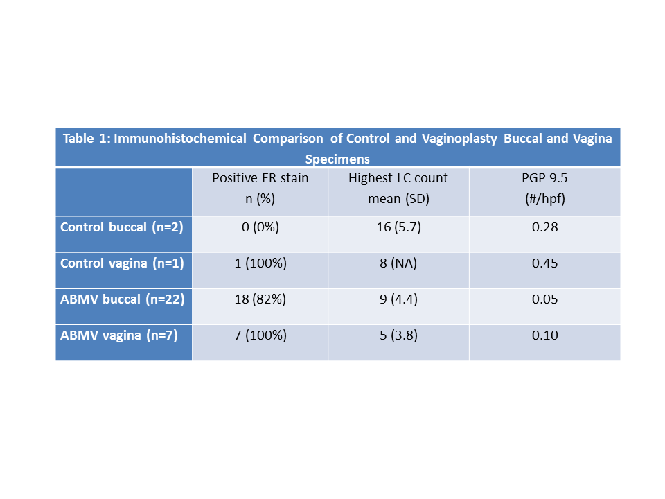
Results: Of 44 patients who had ABMV, seven 46,XX post-pubertal patients who underwent a paired buccal and vaginal biopsy were analyzed, generating 29 biopsies (7 native vagina, 22 buccal graft) at a mean age of 18.8 years (12-27). 9 early and 13 late buccal biopsies were performed. ABMV indication was vaginoplasty stricture repair (2 cloaca, 2 CAH, 1 vaginal agenesis) and distal vaginal augmentation (1 vaginal rhabdomyosarcoma, 1 vaginal agenesis). All native vaginal and a majority of buccal specimens had positive ER staining with no difference between the two, (p=0.546). Buccal specimens were more likely to be only focally or partially positive (i.e. stroma positive, mucosa negative). Mean peak LC count was higher in buccal versus native vaginal specimens (p=0.049). Nerve density/hpf was similar between both tissue types (p=0.5251). Comparing early and late buccal biopsies, there was no difference in positive ER stains (p=0.616), peak LC count (p=0.795), or nerves/hpf (p=0.2276). Comparing
control and ABMV specimens, buccal biopsies had twice the LCs density and half the nerve density of vaginal specimens, Table 1. Control buccal biopsies had 0% while ABMV buccal specimens had 82% ER positivity.
Conclusions: Buccal biopsies revealed conversion from negative to positive ER staining after AMBV grafting. The ratio of nerve and LC densities in buccal versus vaginal specimens were similar in control and ABMV patients. These results may have significant impact on patient counseling regarding future sexual function, risk of STIs, and use of hormonal therapies.

Back to 2014 Fall Congress Meeting Abstracts
|


