
A Rabbit Model for Physeal Function in Bladder Exstrophy: Resection of the Pubic Symphysis Leads to Progressive Deformation of the Pelvic Skeleton
Walter Klyce, BA, Ethan Cottrill, MS, Derek T. Nhan, BS, Casey Kissel, DVM, Caroline Garrett, DVM, Nikolai Sopko, MD PhD, Matthew Kasprenski, MD, Heather Di Carlo, MD, Paul D. Sponseller, MD MBA.
Johns Hopkins University, Baltimore, MD, USA.
Background
Bladder exstrophy typically requires correction of bony anatomy to facilitate successful soft tissue repair. However, the underlying pathogenesis of bladder exstrophy remains elusive. Recent theories suggest that failed closure of the pelvic ring may be the inciting incident. The purpose of this animal study was to examine the role of the pubic symphysis in guiding growth, with the hypothesis that intact symphyseal ligaments are necessary for normal development of the pelvic skeleton.
Methods
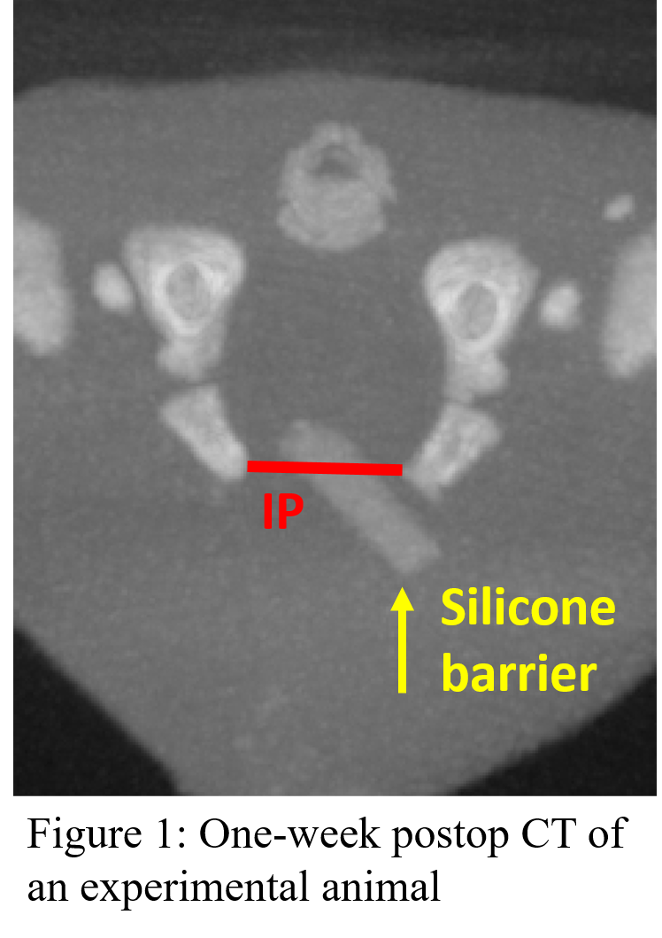
A single investigator performed surgery on 13 rabbits in the first week of life. Nine underwent midline transection of the pubic symphysis and placement a custom silicone implant (1x5x8 mm) to prevent re-fusion of the symphysis. Implants were affixed to the right pubis by nonabsorbable sutures. Four underwent sham surgery. All kits received pelvic CTs (75-micron voxels) at 1 and 5 weeks postoperatively. ImageJ software was used to measure the absolute inter-pubic (IP) distance, inter-ischial (IS) distance, pubic rami lengths, and anterior segment angles. IS/IP ratios and ischiopubic divergence angles, two measures of ischiopubic rotation, were also obtained. Post-euthanasia dissection and caliper-measurement were used to confirm CT accuracy on pre-experiment litters.
Results
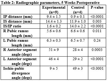
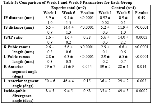
Measurements for week-1 postop CTs are shown in Table 1. Relative to controls, experimental rabbits’ diastasis had increased by mean 3mm one week after surgery. IS and bilateral anterior segment angles also increased significantly after symphyseal transection, though the right anterior segment angle was larger than the left (59±7 vs. 50±6degs, p=0.005). By week 5 (Table 2), the right pubis of the experimental group was shorter than either the contralateral pubis (5.6±0.6 vs. 6.3±0.3mm, p=0.009) or its control-group counterpart (2.6±0.3 vs. 2.9±0.1mm, p=0.025). Change-over-time data is shown in Table 3. Absolute diastasis increased more than twofold (3.9±1.0mm IP vs. 9.4±1.5mm IP, p<0.0001), and rotational measurements (IS/IP and ischiopubic divergence angle) did not return to control values.
Conclusions
In this study we present a novel model for bladder exstrophy in newborn rabbits. To our knowledge, this is the first study to examine the role of the pubic symphysis in guiding pelvic growth. The differences seen in week 1 CTs suggest that this procedure successfully created a pubic diastasis. Chronologic increases in absolute diastasis suggest that the symphysis may play a role in preventing bladder exstrophy in-utero, and the failure of rotational correction hints at a role in guiding pelvic directional growth, as well. 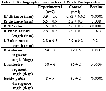
Back to 2018 Program


