
Development and large animal testing of a self-expanding vesicoamniotic shunt and delivery device for fetal LUTO
Michael P. Kurtz, MD, MPH, Kyle Costa, BS, Xuehui Yang, MS, Arthur Nedder, DVM, Alan B. Retik, MD, Caleb P. Nelson, MD, MPH.
Boston Children's Hospital, Boston, MA, USA.
BACKGROUND: Vesicoamniotic shunts (VAS) for fetal lower urinary tract obstruction (LUTO) are associated with a two-fold increase in perinatal survival. The only available devices are plastic double-pigtail shunts developed >30 years ago with major shortcomings: they dislodge in over half of cases and occlusion is common (<=1mm internal diameter). Furthermore, deployment requires antegrade loading, followed by insertion and withdrawal of two separate advancing tools in quick succession. In this study we had two primary objectives: 1) design and manufacture a novel integrated delivery system and VAS, and 2) validate both device and delivery system in a large animal model of fetal LUTO. We hypothesized that the new device would be feasible and practical to deploy, that the VAS would maintain appropriate position without dislodging, would remain patent, and that tissue ingrowth would not occur.
METHODS: The VAS is a self-expanding, lumen-apposing silicone-covered nitinol shunt (Figure 1). The VAS is delivered collapsed to a very narrow diameter, and upon deployment the nitinol mesh expands creating a larger lumen with barbell-shaped retention segments. The delivery device is integrated with its puncture system. For in vivo testing, we developed a fetal Dorset lamb model of LUTO with surgical occlusion of the urachus and bladder outlet at approximately 90 days gestation. 4-14 days later, VAS was placed into the obstructed fetal bladder. The gestation was continued to 130 days (term 145d). Fetal anatomy was examined for VAS position, patency, and tissue ingrowth. Fetal bladders and kidneys were harvested, and stained with Mason's trichrome.
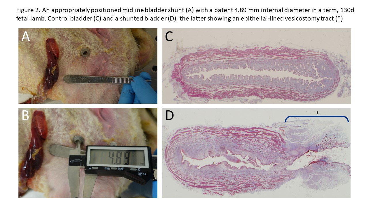
RESULTS: The VAS and delivery device were manufactured in consultation with outside firms. In final configuration, the outer diameter of the delivery device was 2.38 mm, compared with 3.3 mm for the Rocket system and 1.8mm for the Harrison. 5 lambs had shunts placed - two were malpositioned, and all three animals with correctly placed shunts survived to term. At sacrifice, all shunts remained firmly in appropriate position, with withdrawal force between 3 and 3.5 newtons (>303 grams suspended vertically). All VAS lumens were patent with no cases of bladder prolapse or juxta-luminal herniation. No VAS demonstrated tissue ingrowth. Inner diameter of our VAS lumen was 4.9mm (compared to 1mm for the Rocket shunt). There was no fibrosis of the shunted bladders, and the shunts created an epithelium-lined vesicostomy tract (Figure 2). There was no difference in total body weight or lung weight between shunted and non-shunted twin lambs.
CONCLUSIONS: In preclinical large animal testing, the delivery system was able to deploy the VAS in good position. This VAS resists dislodgment, provides a much larger internal diameter for drainage, and is well-tolerated by fetal tissues. It has the potential to improve outcomes by addressing many of the shortcomings of available VAS devices. 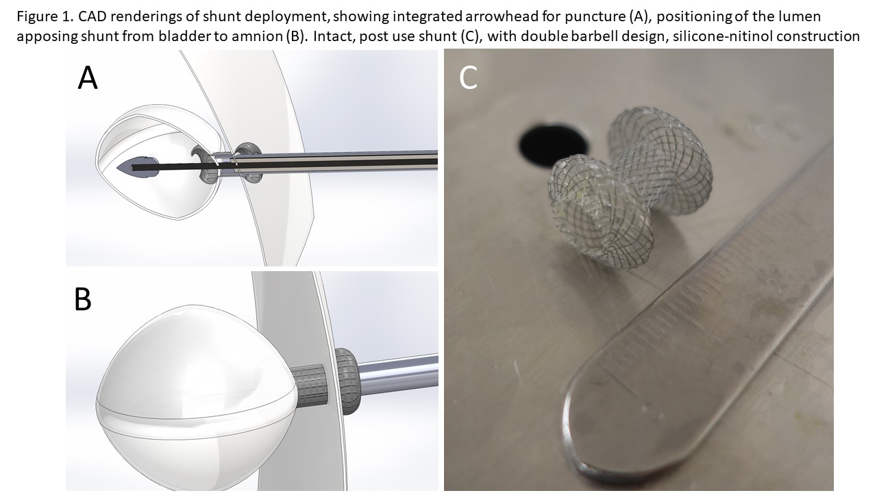
Back to 2019 Abstracts


