
Automated machine learning-driven renal ultrasound interpretation accurately predicts future surgical intervention in patients with prenatal hydronephrosis
Lauren Erdman, MSc, Mandy Rickard, MN, NP, Marta Skreta, BSc, Carson McLean, BSc, Anne-Sophie Blais, MD, Aziz Mezlini, PhD, Michael Brudno, PhD, Anna Goldenberg, PhD, Armando J. Lorenzo, MD.
SickKids, Toronto, ON, Canada.
Introduction:
Predicting surgical intervention for prenatal hydronephrosis (PHN) patients using only ultrasound (US) images provides obvious benefits to this population, namely reduction of invasive testing and faster access to interventions. Herein we describe the development of an automated machine learning approach for prediction of surgery using US images alone for infants with PHN.
Methods:
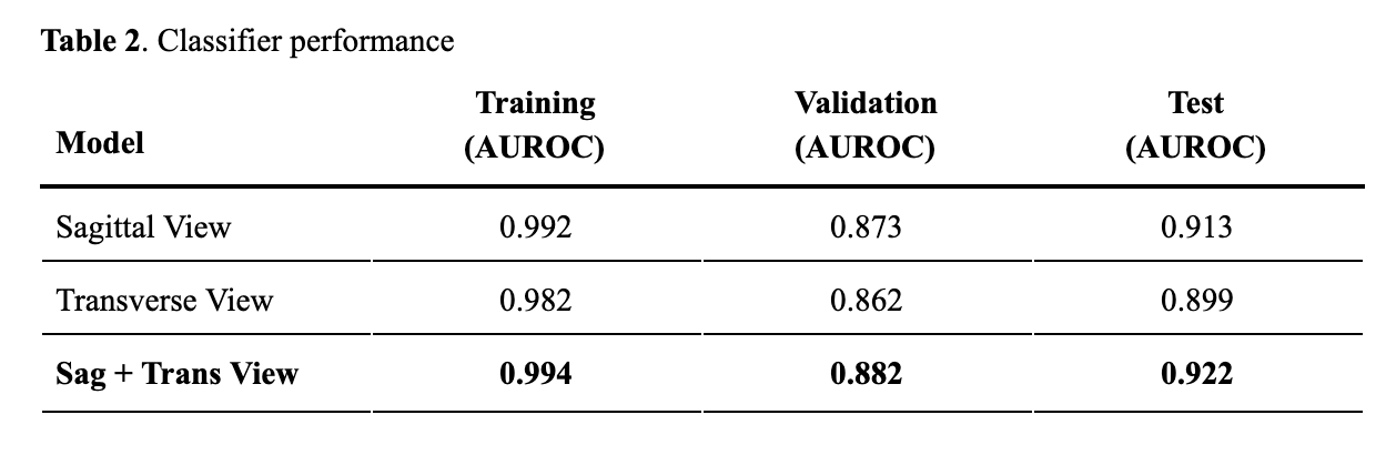
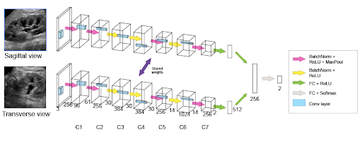
Serial imaging for 205 PHN infants (1359 kidneys) were divided into a training/validation set across 5 folds and each fold was trained independently. A held-out test set (89 individuals, 286 kidneys) was generated with the most recent cases in order to evaluate our algorithm's performance on hypothetical future patients. Our predictive models were convolutional neural networks (CNNs) with the task of predicting surgery from either sagittal or transverse images. To consider information from one view, we tested a CNN with 7 convolutional layers and 2 linear layers. We also investigated the predictive gain from considering both sagittal and transverse views for a given kidney simultaneously. We used a Siamese neural network (SiamNet) composed of two identical CNN subnetworks (Figure 1) that took in as input images from both views to make a prediction.
Results:
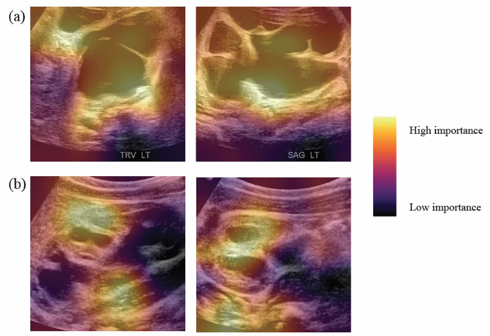
Patient characteristics can be reviewed in Table 1. Including both sagittal and transverse views in the model improved our performance on a held-out test set with an AUROC of 0.922 vs. sagittal (0.913) or transverse (0.899) only (Table 2). With a 5% false negative rate in our surgical cases, our algorithm accurately identifies over half (53%) of non-surgical PHN cases with high confidence. Of these non-surgical cases, 58% of them can be identified on their first ultrasound and 87% by their second ultrasound. This early identification is notable as 25% of those we identify as non-surgical in their first two ultrasounds go on to receive a renal scan. To improve the interpretability of our predictions, we generated heat maps to view areas of interest in US images that our classifier deemed most indicative for predicting surgery (Figure 2a) or no surgery (Figure 2b).
Conclusion:
Surgical prediction for PHN based on US images alone appears to be feasible. Our classifier accurately discriminates between surgical and non-surgical kidneys without feature-engineering or clinical/patient variables from baseline or early ultrasounds. This technology may allow for closer monitoring of infants likely to undergo surgery and reduce exposure to invasive testing for those most likely to resolve. 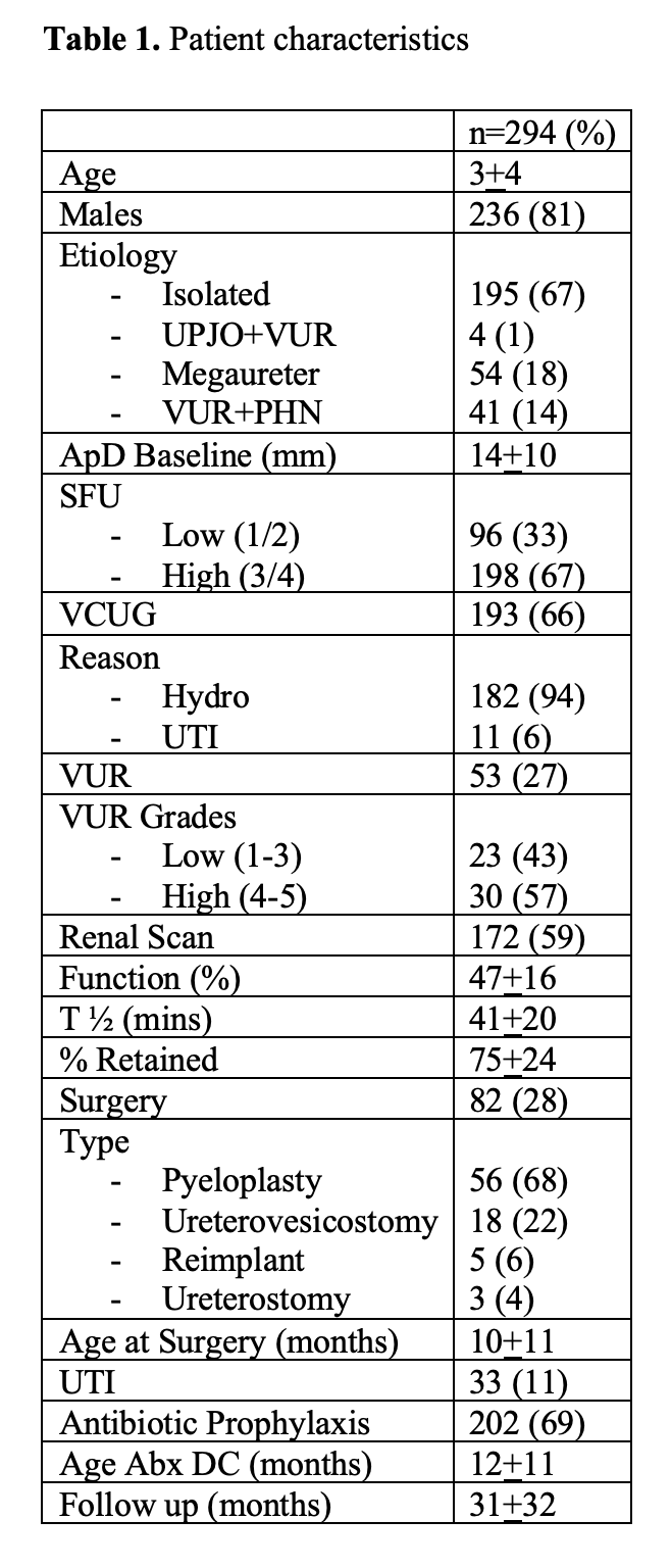
Back to 2019 Abstracts


