
MRI Connectivity Analysis Provides Evidence of CNS Mode of Action for pTENS- A Pilot Study
Jose Murillo Netto, MD, Ph.D1, Dustin Scheinost, Ph.D2, John Onofrey, Ph.D2, Israel Franco, MD2.
1Universidade Federal de Juiz de Fora, Juiz de Fera, Brazil, 2Yale University, New Haven, CT, USA.
Introduction:Parasacral transcutaneous electrical nerve stimulation (pTENS) is a common treatment modality for patients with overactive bladder (OAB). Its mechanism of effectiveness has yet to be fully elucidated. Recent work with fMRI in adults with implanted sacral nerve stimulators imputes its effectiveness on changes in the brain involving the frontal areas. A most recent study using pet scanning showed similar results further strenthening our understanding of how pTENS may work. None of these prior studies utilized sham stimulation to verify that the location of any stimulation is not ubiquitous to the frontal lobes. Our aim was to evaluate MRI functional connectivity analysis to define where in the brain pTENS produces its effects utilizing a sham control stimulation.
Methods: 10 adult volunteers without urinary tract symptoms as assessed by OABSS questionnaire underwent fMRI. Electrodes were placed on skin at sacral level (S2) (pTENS) and on the right scapular region (Sham Stimulation - sTENS). Stimulation was done in each site for 6 minutes at a frequency of 10 Hz and pulse width of 260 μs and intensity determined by the motor threshold. A 6 minutes resting state fMRI was also done as control. Functional connectivity data was acquired during each state (resting, pTENS and sTENS). Standard functional connectivity preprocessing was performed. Seed connectivity was examined to investigate changes in ACC functional connectivity between the stimulations and resting-state conditions. Significance was assessed at p<0.05 corrected for multiple comparisons.
Results:For all conditions (pTENS, sTENS, and rest), standard patterns of ACC connectivity were detectable with strong connectivity between the ACC and subcortical regions and between the ACC and the frontal lobe. Functional connectivity between ACC seed and the dorsal lateral prefrontal cortex (DLPFC) was significantly increased during pTENS compared to rest. sTENS did not increase connectivity between the ACC seed and DLPFC when compared to rest.
Conclusions:Preliminary results indicate that ACC is a major site of activation during pTENS. The ACC is considered the interoceptive motor cortex and is responsible for increased sympathetic activation of the bladder while the DLPFC is responsible for Parasympathetic of the bladder. Increased connectivity between ACC and DLPFC may be a possible mechanism of pTENS effectiveness allowing for proper balancing of sympathetic and parasympathetic activity to the bladder. These connectivity findings appear to be specific to pTENS compared to sTENS. Future work will be to follow this pilot study with more extensive work in patients with OAB. 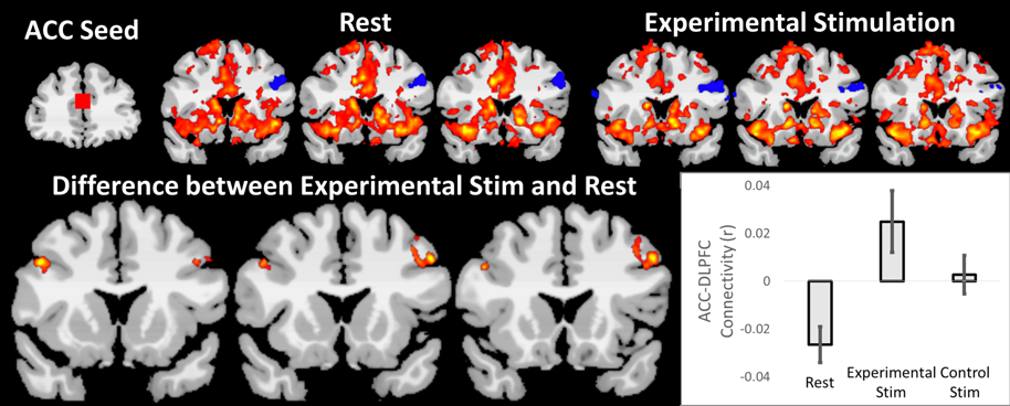
Back to 2019 Abstracts


