
Androgen and Estrogen Receptor Expression in the Developing Human Penis and Clitoris
Yi Li, MD1, Adriane Sinclair, pHD1, Maya Overland, MD, pHD1, Dylan Isaacson, MD2, Gerald Cunha, pHD1, Laurence Baskin, MD1.
1UCSF, San Francisco, CA, USA, 2UCLA, Los Angeles, CA, USA.
Background:
Recent evidence suggests that estrogens along with androgens are necessary for male sexual differentiation, augmenting Jost's original hypothesis. To better understand the mechanism of action of androgens and estrogens in human genital development, we sought to define the precise location of the androgen receptor (AR), estrogen receptors alpha (ERα) and beta (ERβ) in the developing human penis and clitoris.
Methods:
Thirty-six human male and female fetal genital tubercle specimens, 8 to 17-week gestation were processed for scanning electron microscopy, optical projection tomography and immunohistochemical staining. Transverse and sagittal sections six microns in thickness were immunostained with antibodies to AR, ERα and ERβ.
Results:
Scanning electron microscopy of the developing human fetal penis and human clitoris reveals a divergence in genital morphology after the indifferent stage (8 weeks). Canalization of the urethral plate in males and vestibular plate in female occurs in both sexes, but fusion of the urethral folds to form a penile urethra and formation of a glanular urethra only occurs in males (9-17 weeks) (Figure 1).
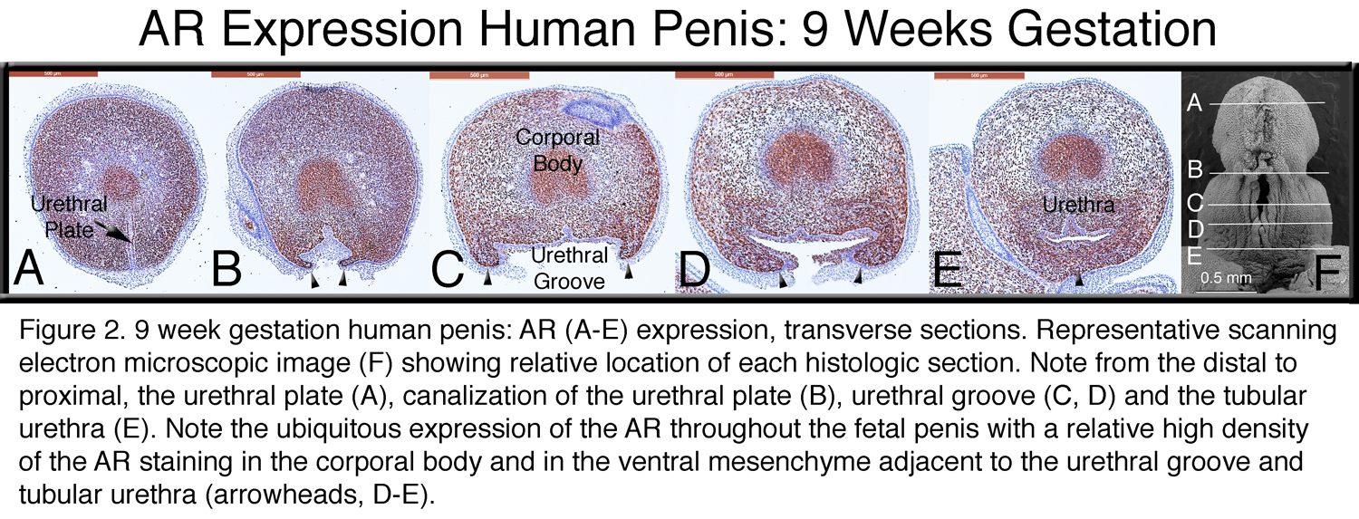
AR is expressed in the developing penis in (a) the mesenchyme lateral to the urethral groove, (b) the surrounding mesenchyme of the newly formed tubular urethra (Figure 2) and (c) in the area of canalization, remodeling and formation of the glanular urethra.
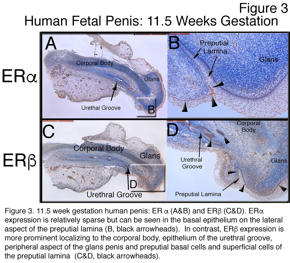
Overall, ERα expression was less prominent than ERβ expression in both the developing penis (Figure 3) and clitoris. ERα expression specifically localized to the epithelial cells of the preputial lamina (Figure 3B) and in the area of canalization and remodeling of the glanular urethral plate. In contrast, ERβ expression was more prominent in (a) the corporal body and the glans mesenchyme (Figure 3C&D), (b) the urethra/vestibular epithelium, (c) the preputial lamina and (d) the glanular urethra.
Conclusions:
AR, ERα and ERβ are all expressed to varying degrees and in specific locations in the developing penis and clitoris. The location of the AR expression is in close proximity to the urethral groove and in the area of formation of the penile and glanular urethra supporting the role of androgens in normal male urethral fold fusion and development. The expression of ERα and ERβ in the epithelial cells of the preputial lamina and in the area canalization and remodeling of the glanular urethral plate is consistent with the possible role of estrogens in normal penile development. Our data is also consistent with the hypothesis that disruption of the AR, ERα and ERβ can lead to impaired virilization of the penis and hypospadias by affecting fusion of the penile urethral folds and remodeling of the glanular urethra. 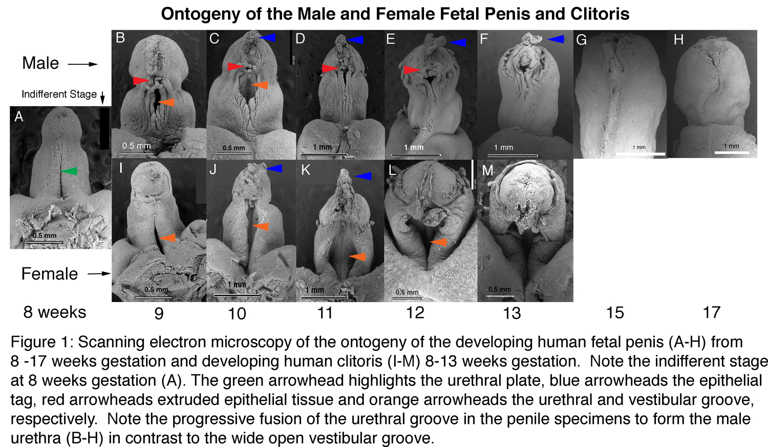
Back to 2019 Abstracts


