
Quantifying the force needed for ureteral stent removal: Initial evaluation of a magnetic stent removal device on benchtop and porcine models
Kunj R. Sheth, MD, Jeffrey T. White, MD, Kathleen Puttmann, MD, David Waters, MD, Matias Soto, MD, Martin Bell, MD, Tasha Aboufadel, MD, Michael J. Heffernan, MD, Eric Richardson, MD, Sang Hoon Song, MD., Ph.D., Chester J. Koh, MD.
Division of Pediatric Urology, Department of Surgery, Texas Children's Hospital/Scott Department of Urology at Baylor College of Medicine, Houston, TX, USA.
BACKGROUND: Indwelling ureteral stents are commonly used in pediatric surgeries for kidney stones and upper tract urinary obstruction, but often require instrumentation or anesthesia for removal. We are evaluating the use of external and catheter tip magnets to remove indwelling ureteral stents with a distally attached magnetic bead. Since little is known about the forces required to remove these stents, our objective was to characterize and quantify the required forces for stent removal for future prototype testing.
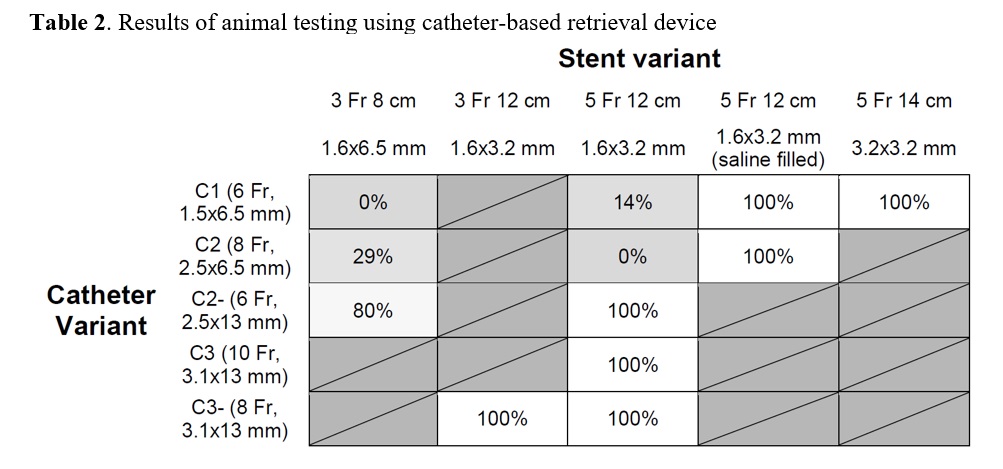
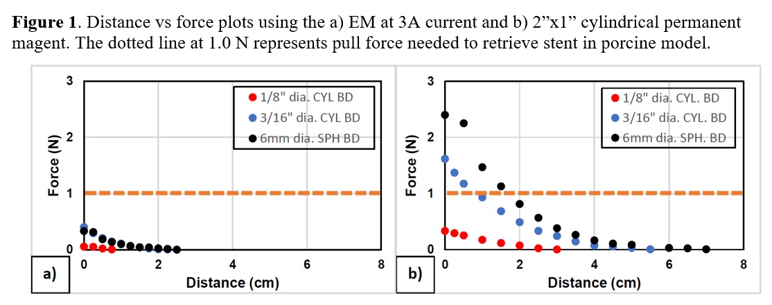
METHODS: A Lazarus 3D urinary tract model was used for benchtop testing, and after obtaining IACUC approval 6 female pig studies took place over a 9-month period. To better emulate the human female urethral anatomy a modified porcine urethral model was created by ligated the urethra distally and tunneling it under the skin to a neomeatus with about 4.5cm urethral length. Using the HF-10 digital force gauge (M&A Instruments), we quantified the force required to remove a variety of different stents (size, presence of curl, and distal magnetic bead). These force measurements were compared to the quantified magnetic force generated by external magnets and catheter tip magnets. Furthermore, the magnetic retrieval devices were tested with various magnetic beads on both benchtop and porcine models. RESULTS: The required force for removal of a 5Fr x 14cm double-J stent was significantly higher in the benchtop model compared to the pig model (4.7N v. 0.8N, p<0.001, Table 1). The external magnets would need to generate the required force of 1N across a 4-5 cm distance from the urethral meatus to the bladder neck. Figure 1 shows distance versus force plots for the strongest external permanent magnets and electromagnets. The force generated at a distance of 4-5cm was orders of magnitude lower than the necessary force, and thus they failed to retrieve the magnetic bead stent in the bladder. The catheter retrieval device showed better success with a variety of different retrieval magnet and stent bead sizes (Table 2). The addition of saline to the bladder allowed for better retrieval rates of the smallest beads (1.6 x 3.2mm), even by the smallest magnetic tip catheters (1.5 x 6.5mm)
CONCLUSIONS: The force required for ureteral stent removal is under 1N on the porcine model, and improved benchtop models that emulate such parameters will facilitate future testing. The external magnets could not generate sufficient force due to its inverse square variation with urethral distance. In contrast, the catheter tip magnet model overcomes the limitation of distance. Further studies are needed to define the optimal combination of catheter magnetic tip size and stent magnetic bead size. 
Back to 2019 Abstracts


