
Flat panel detector c-arms are associated with a dramatic reduction in absorbed radiation dose during ureteroscopy
Michael P. Kurtz, MD, MPH, Bartley G. Cilento, Jr., MD, MPH, Michael G. Demers, AS RT(R), Robert D. MacDougall, M.Sc, Ph.D., Ian R. McCarthy, BA, Alyssia M. Venna, MBS, Caleb P. Nelson, MD, MPH.
Boston Children's Hospital, Boston, MA, USA.
BACKGROUND: Pediatric urologists treating urolithiasis aim to achieve excellent surgical results with the lowest reasonably achievable radiation dose. A recent advance in fluoroscope technology, the change from analog image intensifiers to digital flat panel detectors (FPD), has the potential to reduce dose during image-guided procedures. We wished to study the radiation doses during the gradual introduction of FPD C-arm fluoroscopes in a single pediatric institution, hypothesizing that FPDs would be associated with a substantially lower dose than from fluoroscopes with image intensifiers.
METHODS: We reviewed retrospectively a consecutive series of retrograde ureteroscopy cases for urolithiasis at a single institution. FPDs were introduced slowly, and assignment of FPD or traditional image intensifier flouroscope was dictated by OR demands, or the procedure location at a satellite at which an FPD was not available. We restricted to our analysis one year's worth of cases, targeting the maximal overlap between the two fluoroscopes to limit the effect of environmental influences on the procedure. We collected procedural, demographic, and dose information. Flouroscope settings, positioning, and collimation strategies were standardized with a separate fluoroscopy timeout at the start of all procedures.
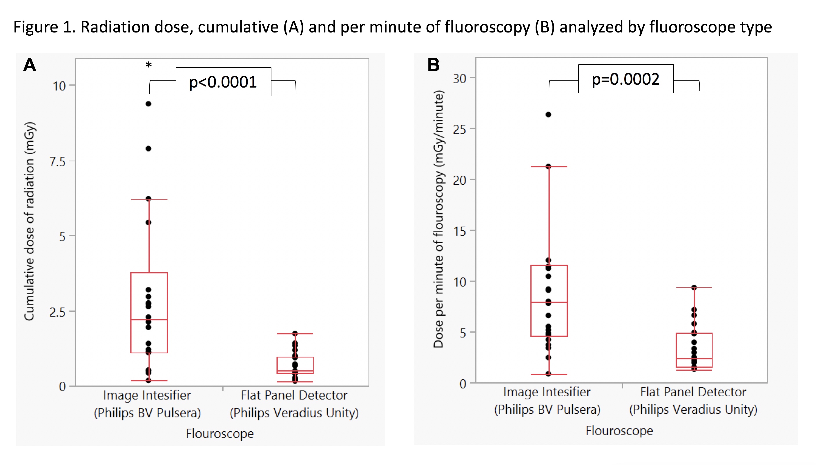
RESULTS: A total of 44 unique patients were treated with retrograde ureteroscopy between January 2018 and January 2019. Characteristics did not significantly differ between groups with respect patient factors, measures of urolithiasis size or location, or procedural complexity (Table 1). The median dose was over four times higher in the image intensifier group than in the flat panel detector group, median 2.2 [1.1, 38] mGy versus 0.48 [0.42, 0.97], p<0.0001 (Figure 1). The highest dose in the FPD group was 1.73 mGy, lower than median dose in the image intensifier group. This dose was over 3-fold higher correcting for fluoroscopy time, and remained significant in a multivariate model including fluoroscopy time and patient weight (β=2.4, p=0.007).
CONCLUSIONS: This is the first report of radiation dose from c-arms comparing image intensifiers and FPDs in adult or children. Use of an FPD was associated with a substantial decrease absorbed dose for patients undergoing ureteroscopy. Flouroscopy equipment considerations should be integrated into comprehensive ALARA strategies for urolithiasis treatment. 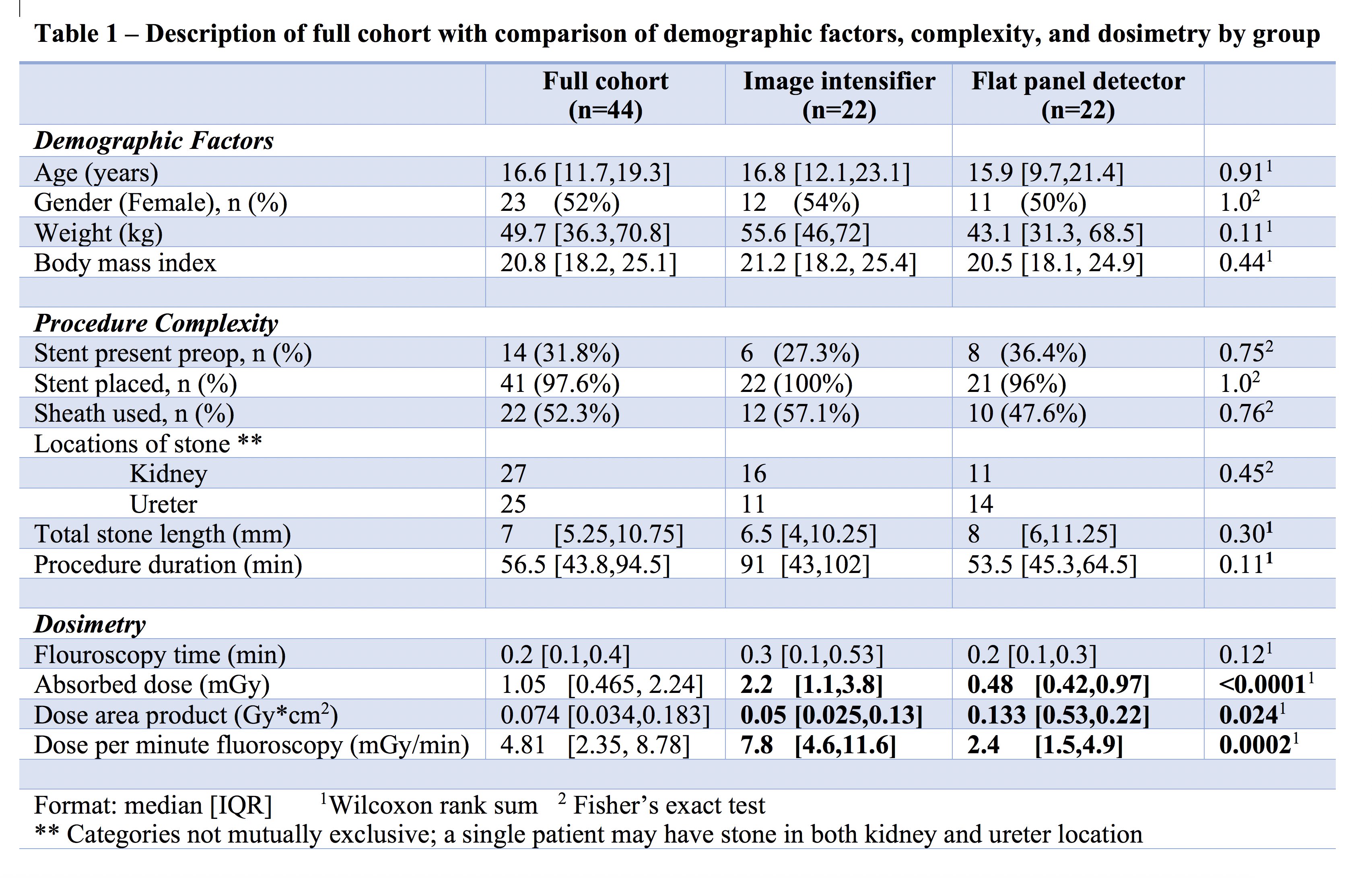
Back to 2019 Abstracts


