
A new appendicostomy technique to prevent stomal stenosis
Eric A. Kurzrock, M.D..
UC Davis Children's Hospital, Sacramento, CA, USA.
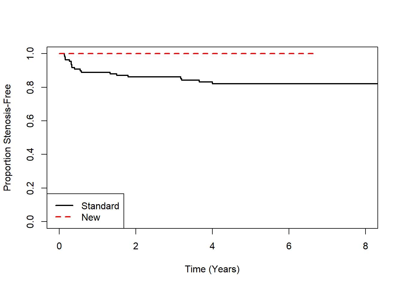
Background Stomal stenosis has been reported to occur in 10 to 40% of patients after appendicostomy for ACE Malone and continent urinary diversion. The standard stoma technique entails excision of the distal appendix, antimesenteric spatulation and anastomosis to a flap of skin. We hypothesized that extensive excision of the firm posterior umbilical skin, preservation of the appendiceal tip and accessing the lumen in a more proximal and vascular area would lessen the incidence of stenosis. Methods The records of patients who underwent ACE Malone or urinary diversion with appendix between 2000 and 2018 were reviewed. Patients who had a split appendix technique or less than one year of follow up were excluded. The new appendicostomy (Figure) was implemented in 2012. The incidence of patient, surgical and anatomical variables such as age, gender, obesity, open or laparoscopic approach, ACE or urine diversion, cecal or appendiceal adhesions, retrocecal appendix, imbrication or tunneling and stomal stenosis were recorded for the two cohorts and compared with Wilcoxon rank sum tests for continuous variables and Fisher's Exact Test for categorical variables. Time to stenosis was modeled by patient and surgery characteristics using Cox proportional hazards models. Results Inclusion criteria were met by 138 patients, 30 newer stomas and 108 standard stomas. Median follow up after the new and standard techniques was 3 and 9 years, respectively. Beyond follow up time, the two cohorts had no significant difference in covariates. Stomal stenosis occurred in 19 of 138 (14%) patients, 5 of 26 urine diversions and 14 of 112 ACE patients. All were revised and continue to be used. Stenosis has not occurred in 30 patients with the new stoma technique, hazard ratio was 0.103 (CI 0.000811, 0.755) (P < 0.02) (Chart: Kaplan-Meier plot of time to stenosis). No other patient, anatomical or surgical variable was associated with stenosis. Conclusions A more extensive excision of the posterior umbilicus to isolate healthy skin, preservation of the appendiceal tip and vasculature and accessing the lumen more proximal are easy modifications to appendicostomy that appear to lessen the risk of stenosis. 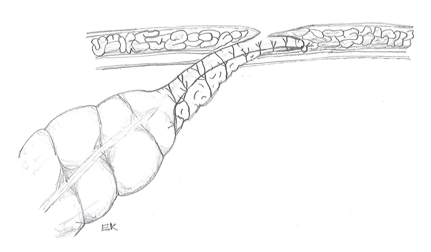
Back to 2019 Abstracts


