
Genital Anatomy of Female Pediatric Patients With Genital Cutting: A MRI Based Cross Sectional Study
Avi Baskin, MD1, Pinar Karakas-Rothey, MD2, Laurence Baskin, MD1.
1UCSF, San Francisco, CA, USA, 2UCSF Benioff Children's Hospital of Oakland, Oakland, CA, USA.
Background:
Female Genital Cutting (FGC) is defined by the World Health Organization (WHO) as all procedures involving partial or total removal of the external female genitalia for ceremonial or non-medical reasons. There are four types of FGC classified by the WHO that range from partial removal of the clitoris and prepuce to a complete narrowing of the vaginal orifice and clitorectomy. There are no studies that describe radiologic anatomy of this group of pediatric patients. The aim of the study is to describe the radiologic anatomy in a subset of this group of patients. We hypothesize that patients who underwent this type of female genital cutting would have anatomical injury to the clitoris and decreased clitoral body width and height.
Methods:
This is a case control study of sexual anatomy in five pediatric patients at a single institution who underwent type 1 (partial removal of the prepuce) FGC. The anatomy of the clitoris and bulbs was examined with pelvic MRI on 3Tesla Philips Achieva, Philips Medical System, Netherlands. We used aged matched controls for a comparison group and measured the female external genitalia structures on pelvic MRIs (on 3T and 1.5T Philips scanners) that were performed for other indications. To limit the number of comparisons we averaged the size structures that were bilateral. We used t-testing to compare measurements between the girls with and without female genital cutting.
Results:
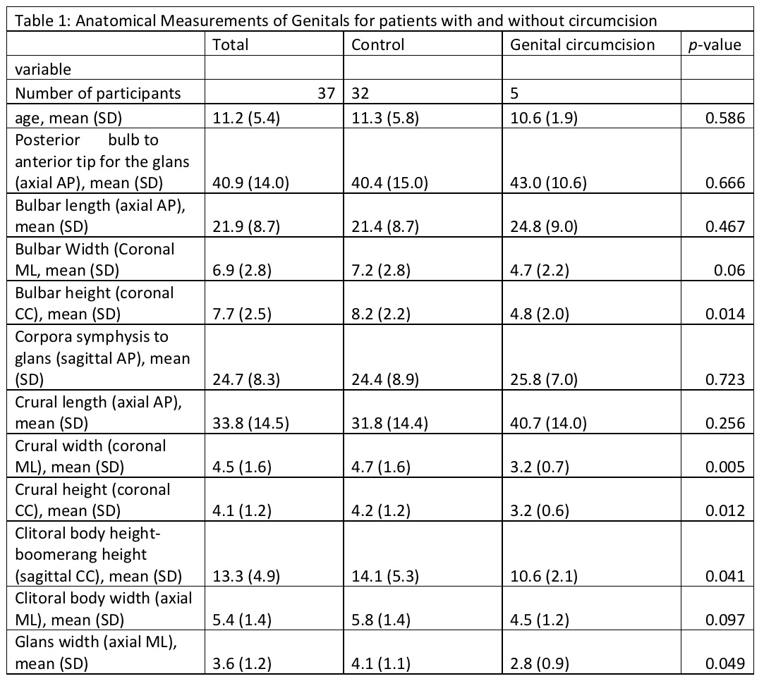
There were 37 total participants in the study. Five patients had undergone FGC and 32 patients were controls. The average age in the group who underwent female genital cutting was 11.2 years which was similar to the average of 11.3 years in the control group (p=0.58). There was no evidence of anatomical injury to the glans clitoris, clitoral body and crus between patients who underwent FGC and controls. Table 1 highlights comparisons of anatomical size between patients. Figure 1 shows a selection of sample MRI images.
Conclusion:
MRI was a useful tool to define anatomy of the female genitalia. Patients subject to type 1 FGC showed grossly normal anatomy with no evidence of injury to clitoral body or glans. This information is important to counsel patients and families who present to health care after FGC. There is statistically significant decreased size of crural width and height as well as glans width. Further study and follow up is needed to reveal if these statistically significant changes are maintained and clinically relevant as these patients age. 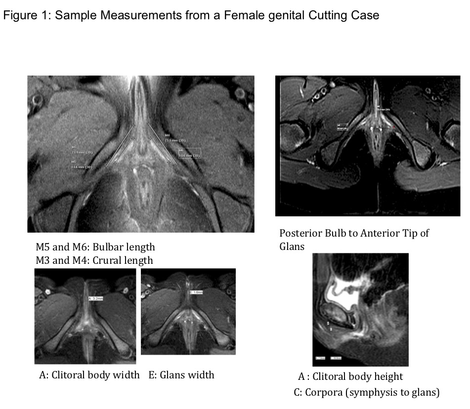
Back to 2019 Abstracts


