
Super Resolution Structured Illumination Microscopy: A Novel Three-Dimensional Technique To Study Mitochondrial Networks in the Urinary Bladder
Ching Man Carmen Tong, DO1, Stacy T. Tanaka, MD1, John C. Thomas, MD1, John C. Pope, IV, MD1, Abby S. Taylor, MD1, Mark C. Adams, MD1, John W. Brock, III, MD1, Volker H. Haase, MD2, Douglass B. Clayton, MD1.
1Division of Pediatric Urology, Monroe Carell Jr. Children's Hospital at Vanderbilt, Nashville, TN, USA, 2Department of Medicine, Vanderbilt University Medical Center, Nashville, TN, USA.
Introduction
The mitochondrion plays a vital role in cell metabolism and energy production. While it has been extensively studied in renal cancer and injury, the role of mitochondrial dysfunction in the bladder remains unclear. Recent efforts have begun to focus on the role of mitochondria as potential therapeutic targets and disease markers in bladder cancer. Super resolution structured illumination microscopy (SIM) is a novel technique that uses patterned illumination to provide three-dimensional, high-frequency spatial information otherwise not producible through conventional imaging. By rendering images that are of twice the resolution of a regular optical microscope, morphological analysis and networks of subcellular structures can be appreciated. Here, we investigate the application and utility of this technique to visualize three-dimensional mitochondrial dynamics in mice bladders.
Methods
All experiments presented were approved by our institutional animal care and use committee. Eight week-old female wild-type C57BL/6 mice were studied. Bladders were distended using formalin after animals were sacrificed, and tissues were fixed and paraffin embedded following standard protocols. Sections of the bladder were mounted on coverslips treated with (3-Aminopropyl)trimethoxysilane and stained for mitochondrial inner and outer membrane markers (COXIV and VDAC, respectively), and then imaged by Nikon N-SIM microscope. Changes in organelle structure were quantified using Imaris software.
Results
SIM imaging demonstrated not only individual mitochondria within each umbrella cell, but also distinguished inner membrane (denoted pink) from outer membrane (denoted green) (fig a). Tight correlations between the inner membrane and the outer membrane can be appreciated by 3D image-rendering with Imaris software (fig b). Mitochondria appear to be more abundant around the nucleus as compared to the cytosol facing luminal surface within each umbrella cell. Utilization of this technique also allows for quantification of mitochondrial volume through measurement of membrane markers (fig c).
Conclusion
SIM imaging is a novel technique that provides three-dimensional visualization and analysis of mitochondrial morphology and dynamics. To our knowledge, this is the first successfully applied SIM technology to bladder tissue and we revealed clear associations between the inner and outer membrane networks of the mitochondria. Future studies using human bladder tissue will help identify the role of mitochondrial dysfunction in various pediatric diseases. 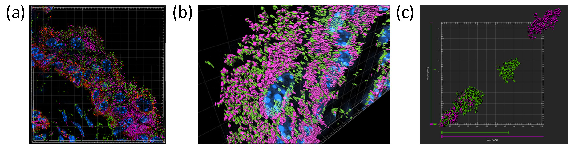
Back to 2019 Abstracts


