
A Case of a Large Abdominal Cyst with Diagnosis of Crossed Fused Ectopic Kidney with Severe UPJ Obstruction
Chris Ballantyne, Marisa Gray, Sean T. Corbett, Nora G. Kern
University of Virginia, Department of Urology
RS is a 5 year old girl who was adopted from India with limited care. She has a history of an ectopic anus s/p repair, bilateral radial hypoplasia, and a right solitary kidney. A history of cystic mass was not known prior. On arrival to the USA, a renal ultrasound was done to evaluate the solitary kidney; the patient was found to have a large midline abdominal cystic mass. She was sent to pediatric surgery to rule out malignancy. An MRU was done which showed the cystic mass abutted the lower/mid-pole of the right kidney, and it appeared there was a rim of renal tissue surrounding part of the cystic structure. The right kidney was also mal-rotated anteriorly. It was suspected that the patient may have a cross fused ectopic kidney with severe UPJ obstruction of the crossed kidney. Due to this, a Mag3 renal scan was obtained which demonstrated no uptake into the large cystic mass and 100% function in the right kidney. The patient was taken to the OR for robotic partial nephrectomy/removal of the cystic mass. Intra-operatively, the large dilated mass was found to be fused to the inferior pole of the right kidney with a large aberrant vessel overlying the junction. An atretic blind ending ureter was found under this vessel. Pathology was consistent with cystic dysplastic changes. The patient did well post-operatively. Repeat renal ultrasound post-surgery demonstrated well healing isthmus tissue; however ultrasound 10 months post-op demonstrated the isthmus tissue appeared more multi-cystic in nature. She continues to be followed.
Images Pre-op: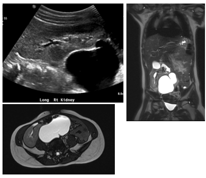
Image Intra-op: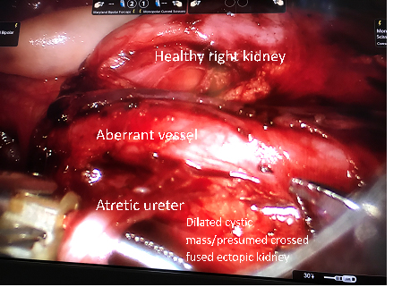
Images Post-op: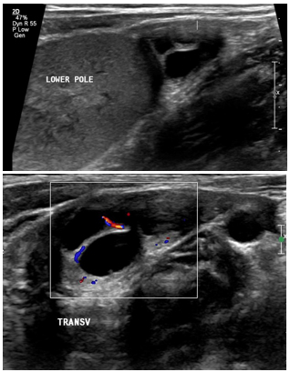
Back to 2019 SFU Program


