Back to 2026 Abstracts
Practice Patterns and Predictors of Varicocele Repair in Adolescents
Samy Sasoun, B.S.1, Sargam Panpaliya, B.S.1, Mia Kotikovski, B.S.1, William T. Berg, M.D.2, Michael Ernst, M.D.2.
1Renaissance School of Medicine at Stony Brook University, Stony Brook, NY, USA, 2Department of Urology, Stony Brook University Hospital, Stony Brook, NY, USA.
BACKGROUND: Varicoceles affect up to 20% of adolescent males and are associated with impaired fertility. Unlike in adults, indications for surgical intervention in pediatric patients remain controversial, with management variability based on testicular asymmetry, pain, and inconsistently obtained semen analyses. We evaluated rates of varicocele repair, utilization of semen analysis, geographic variation, and predictors of intervention in a multi-institutional cohort of adolescent males with varicocele.
METHODS: The TriNetX Research Network was queried to identify males aged 10-20 years diagnosed with varicocele (ICD-10 I86.1). Surgical repair rates within 1- and 6-year follow-up were determined using procedural codes. Demographics, pain-related diagnoses, imaging patterns, semen analysis utilization, and geographic trends were reviewed. Predictors of repair were assessed using a multivariable Cox proportional hazards analysis comparing patients with and without pain diagnoses while adjusting for age, testicular atrophy, and duplex imaging.
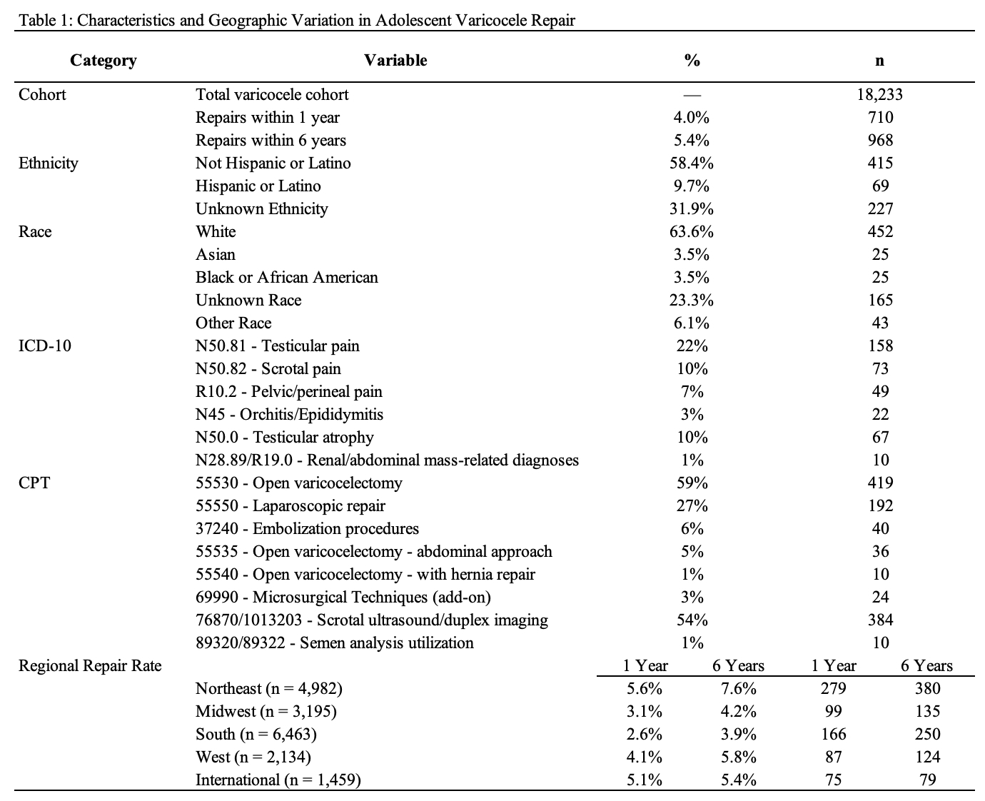
RESULTS: Across 102 healthcare organizations, 18,233 adolescent males with varicocele were identified. Mean age at diagnosis was 14 years, while patients undergoing repair had a mean age of 18 years. Surgical intervention occurred in 710 patients (4.0%) within 1 year of diagnosis and in 968 patients (5.4%) within 6 years.
Geographic variation in repair rates was observed across regions, with the Northeast demonstrating the highest 1-year repair rate (5.6%) and the South the lowest (2.6%). Among patients undergoing repair within 1 year (n = 710), open varicocelectomy was the most common approach (59%), followed by laparoscopic repair (27%), embolization (6%), abdominal approach repair (5%), microsurgical technique add-on coding (3%), and hernia repair (1%). Pain diagnoses, including scrotal, testicular, and pelvic pain, were identified in 30% of patients, while testicular atrophy was identified in 10%. Scrotal ultrasound, including duplex imaging, was performed in 54% of patients. Semen analysis utilization and renal or abdominal mass diagnoses were uncommon (1%) (Table 1).
On multivariable Cox proportional hazards analysis using 6-year follow-up, increasing age (HR 1.086, 95% CI 1.057-1.115, p < 0.001) and duplex imaging (HR 1.594, 95% CI 1.037-2.451, p = 0.034) were associated with repair. Pain diagnoses (HR 0.846, 95% CI 0.716-0.999, p = 0.049) and testicular atrophy (HR 1.222, 95% CI 0.305-4.907, p = 0.777) were not associated with an increased likelihood of repair.
CONCLUSIONS: Adolescent varicocele repair is performed in a minority of patients, with geographic variability in evaluation and management. Increasing age and duplex imaging utilization were associated with a higher likelihood of repair, suggesting that older age and more extensive diagnostic evaluation may contribute to intervention patterns. Northeastern and international institutions demonstrated higher rates of surgical intervention compared to other regions, suggesting regional variation in management thresholds. Low semen analysis utilization highlights inconsistencies in fertility-focused evaluation. These findings underscore a lack of standardized, evidence-based criteria, supporting the need for more uniform clinical guidance in adolescent varicocele management.
Back to 2026 Abstracts


