Back to 2026 Abstracts
Multidisciplinary Approach to Managing Pediatric Genitourinary Vascular Anomalies: Patient Characteristics and Role of the Urologist
Andrew Gabrielson, MD1, Halsie Donaldson, MD2, Ashley Talton, BS1, Gabriel van de Walle, MD2, Carolyn Kiolbasa, RN1, Anthony Mancini, MD1, Duri Yun, MD, MPH1, Sarah Chamlin, MD1, Jennifer Reichek, MD1, Noopur Gangopadhyay, MD1, Shankar Rajeswaran, MD1, Joe Baker, MD1, James Donaldson, MD1, James Rague, MD, MS1, Ilina Rosoklija, MPH1, Emilie Johnson, MD, MPH, FACS1.
1Ann & Robert H. Lurie Children's Hospital of Chicago, Chicago, IL, USA, 2University of Illinois at Chicago College of Medicine, Chicago, IL, USA.
BACKGROUND: Genitourinary (GU) vascular anomalies (VAs) are rare (<5% of VAs overall) and heterogenous, encompassing venous, lymphatic, and mixed malformations that may cause swelling, pain, or functional impairment. This study describes patient characteristics, management patterns, and role of the urologist for children with GU VAs evaluated at a tertiary, multidisciplinary clinic.
METHODS: We conducted a retrospective review of patients with GU VAs seen from 2013-2025. Demographics, lesion type, anatomic distribution, imaging characteristics, and interventions were abstracted. Descriptive statistics were used to summarize patient and disease characteristics, and specialties involved in care.
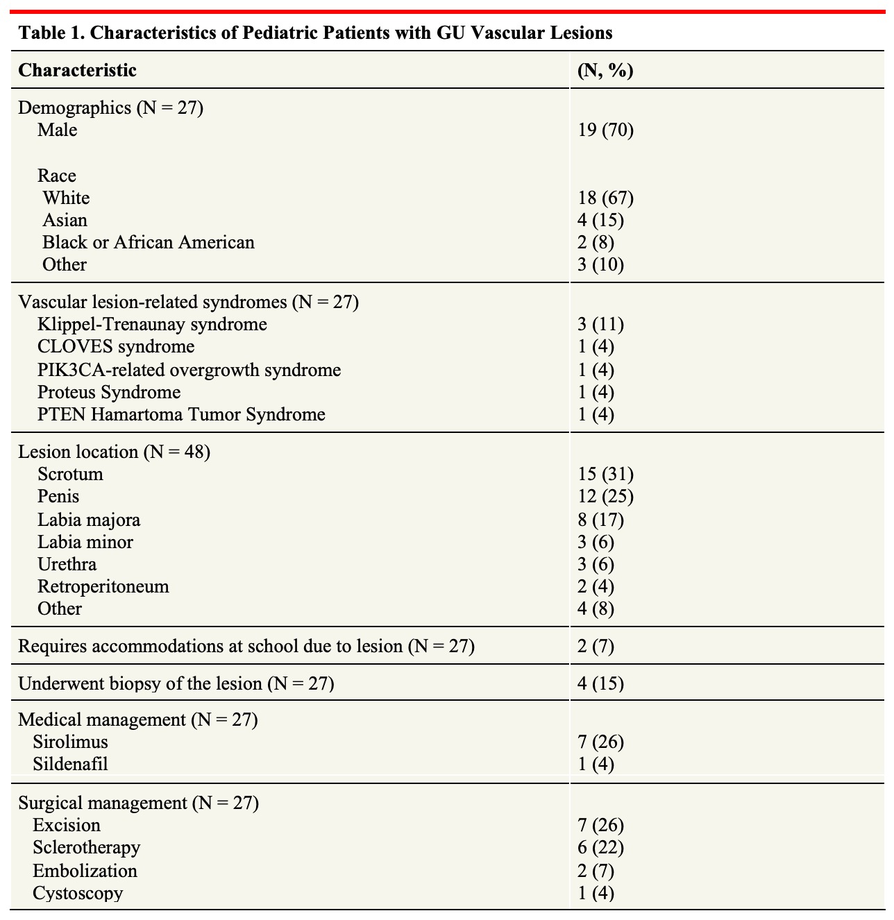
RESULTS: Twenty-seven patients with 48 lesions were identified: 19 (70%) male, median age at diagnosis 2.4 years (range 0-16 years). The most common diagnoses were venous malformation (33%), mixed venolymphatic malformation (22%), or lymphatic malformation (15%). GU VAs were most frequently located on the scrotum (31%), penis (25%) and labia majora (17%); 63% of patients had vascular lesions in non-GU areas. Most patients were asymptomatic (55%), however pain (30%), bleeding (30%), and swelling (19%) were common presentations. Lesions impacted activities of daily living in 29%. Imaging was obtained in all patients: 93% had MRI or MR angiography, 44% had ultrasound. Medical management was pursued in 8 patients: 7 received sirolimus and 1 received sildenafil. GU procedures (biopsy, sclerotherapy, excision, embolization) were performed in 16/27 (59%), with 7/16 (44%) by a urologist. Of patients that had a procedure with curative intent and adequate follow up, 6/16 (38%) had a recurrence. Five had complications: 2 wound dehiscence, 1 bleeding, 1 pain, 1 localized swelling. The most common specialties involved in care were: dermatology (24/27, 89%), urology (18/27, 67%), interventional radiology (16/27, 59%), plastic surgery (10/27, 37%), and child psychiatry (9/27, 33%). Follow-up was recommended for most (22/27, 81%), but only 15/27 (55%) are receiving ongoing care.
CONCLUSIONS: This is the largest series of pediatric GU VAs, which were found to be primarily lymphatic or mixed venolymphatic malformations affecting males in the penis or scrotum. Although many patients opted for surveillance without intervention, surgical excision and sclerotherapy are options in symptomatic patients with lesions limiting activities of daily living. Multidisciplinary collaboration is essential for optimal diagnosis and management of these rare entities.
Back to 2026 Abstracts


