Back to 2026 Abstracts
Diagnostic Challenges and Contributing Factors of the Modified Bosniak Classification in Complex Pediatric Renal Cysts: A Scoping Review of Intermediate-Risk Lesions
Carl Suerte, BS, Alexandra Siegal, MD, Niccolo Passoni, MD.
Baylor College of Medicine, Houston, TX, USA.
BACKGROUND: The modified pediatric Bosniak (mBosniak) classification reliably stratifies cysts at the extremes (Class I benign, Class IV malignant) but performs less reliably for intermediate-risk Class III lesions. The factors contributing to this limitation have not been examined together. This scoping review evaluated mBosniak performance in pediatric Class III lesions and synthesized the imaging features, diagnostic workflows, and clinical factors underlying its reduced reliability.
METHODS: A PRISMA-ScR-compliant search of Medline (OVID), Embase, Web of Science, and Cochrane Library (2000-2025) identified studies on pediatric complex renal cysts. Extractable cohort studies with explicit imaging modality and histopathologic confirmation were quantitatively analyzed. Malignancy rates for mBosniak Class III and IV cysts were calculated with Clopper-Pearson 95% confidence intervals across two pools: Pool A (strict CT-only stratification, n=33 cysts) and Pool B (mixed-modality stratification, n=62 cysts). Case reports and non-extractable cohorts were qualitatively analyzed to synthesize diagnostic workflow patterns and map imaging features across complex cyst diagnoses. Bias was assessed using QUADAS-2 and a reporting-missingness assessment.
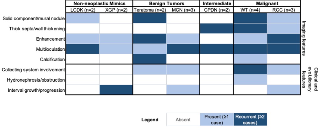
RESULTS: Of 1,204 records screened, 45 studies were included (41 qualitative, 4 quantitative). Class III malignancy rates were 11.1% (95% CI: 0.3-48.3%) in Pool A and 5.9% (95% CI: 0.1-28.7%) in Pool B, notably lower than CT-derived adult benchmarks (44-58%). Class IV malignancy rates were 79.2% (95% CI: 57.9-92.9%) in Pool A and 84.4% (95% CI: 70.5-93.5%) in Pool B, comparable to adult benchmarks (83-92%). Critically, the majority of Class III lesions in both pools represented intermediate-grade pathology (e.g., cystic nephroma, metanephric adenoma) rather than malignancy. Qualitative synthesis of complex cyst case reports revealed a predominantly unidirectional escalation workflow with no observed de-escalation after cross-sectional imaging (0/20 cases). Feature mapping demonstrated substantial overlap in imaging characteristics across benign, intermediate, and malignant entities.
CONCLUSIONS: Three factors explain the limited performance of mBosniak in pediatric Class III lesions: a heterogeneous tumor biology that includes a substantial intermediate-grade pathology component, a rigid escalation-driven workflow favoring surgical management, and non-specific imaging feature overlap across diagnostic categories. The lower Class III malignancy rate does not justify conservative management on its own, as much of this category warrants surgical excision despite being non-malignant. Additional cross-sectional imaging after initial ultrasound characterization may increase diagnostic confidence without improving accuracy. Prospective multi-institutional studies are needed to refine surveillance criteria (incorporating size- and age-based stratification, growth kinetics, and ultrasound feature changes) and to evaluate management against the broader endpoint of pathology warranting surgery, not malignancy alone.
Malignancy rates of mBosniak Class III and IV cysts by imaging classification approach| mBosniak Class | Pool A (Strict CT-Only) | Pool B (Mixed-Modality / Real-World) | Adult Bosniak Malignancy Yield |
| Class III | 1/9 (11.1%); 95% CI: 0.3%-48.3% | 1/17 (5.9%); 95% CI: 0.1%-28.7% | 44%-58% |
| Class IV | 19/24 (79.2%); 95% CI: 57.9%-92.9% | 38/45 (84.4%); 95% CI: 70.5%-93.5% | 83%-92% |
Back to 2026 Abstracts


