Back to 2026 Display Posters
Progression of vesicoureteral reflux in children with posterior urethral valves is associated with adverse short- and medium-term renal outcomes
Adree Khondker, MD, Ali Hamade, MSc, Eviatar Fields, PhD, Samer Maher, BSc, Joana Dos Santos, MD, Michael Chua, MD, Mandy Rickard, MN-NP, Armando Lorenzo, MD.
The Hospital for Sick Children, Toronto, ON, Canada.
BACKGROUND: Identifying patients with posterior urethral valves (PUV) at high risk for renal deterioration is of interest. To date, most predictors have been based on laboratory values. Vesicoureteral reflux (VUR) is frequently identified in patients with PUV and can reflect a combination of bladder outlet obstruction and impaired bladder function, including high-pressure voiding. While some cases of VUR improve following primary valve ablation and bladder decompression, others persist, worsen, or appear later during follow-up. The clinical importance of VUR trajectory remains poorly characterized. We hypothesized that patients with new-onset or worsening VUR would have worse short- and medium-term renal outcomes.
METHODS: We conducted a single-centre retrospective study of patients with PUV who underwent primary valve ablation at our institution between 2002 and 2025 and had two or more VCUGs during follow-up. Patients were excluded if their initial VCUG was unavailable, if they had only a single VCUG, if serial VCUGs were less than 30 days apart, if they underwent upper urinary tract surgery between VCUGs, if the first visit was after 1y of age, or if outcome metrics were missing. Collected data included demographics, imaging, surgical outcomes, and serum creatinine. The primary outcomes were serum creatinine and estimated glomerular filtration rate (eGFR) at 1, 3, and 5 years. Based on VUR status at the index visit and follow-up, patients were grouped as: no VUR at either timepoint (G0), new VUR at follow-up (G1), stable or improved VUR (G2), or worsened VUR (G3). Groups 1 and 3 were considered progressive. Categorical variables were reported as counts and proportions, and continuous variables as median (IQR). Group comparisons used Kruskal-Wallis with post hoc Dunn-Sidak testing.
RESULTS: Of 88 children, 70 had no change in VUR and 18 had progressive VUR. Baseline characteristics were similar between groups, but children with progressive VUR had significantly lower recent eGFR (47 vs 80 mL/min/1.73 m², p=0.019) and higher serum creatinine at 1 year (40 vs 30 μmol/L, p=0.048), 3 years (67 vs 36 μmol/L, p=0.011), and 5 years (85 vs 41 μmol. The between-group difference widened over time (Figure). Across the four VUR trajectory groups, recent eGFR differed significantly (p=0.0009), with the lowest median in Group 1 (36 mL/min/1.73 m²) and the highest in Group 2 (92 mL/min/1.73 m²). Serum creatinine also differed across groups at 1, 3, and 5 years (p=0.045, 0.01, and 0.005, respectively), with Group 1 showing the poorest renal outcomes and creatinine that rose over time.
CONCLUSIONS: New or progressive VUR in patients with PUV is a negative prognostic factor for short- and medium-term renal outcomes. We demonstrated that the subset of patients with worsening VUR on imaging also shows biochemical evidence of decline in kidney function during follow-up. This represents an important, and possibly modifiable, prognostic factor for renal preservation in patients with PUV.
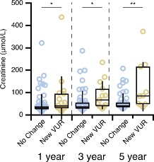
Figure. 1-, 3-and 5-year follow-up creatinine values for children with no change to VUR (groups 0 & 2) and with new-onset/progressive VUR (groups 1 & 3).
Back to 2026 Display Posters


