
Selective release of CO2-loaded nanoparticles for vesicoureteral reflux imaging
Andy Y. Chang, MD1, Helal Syed, MD1, Zoe Nussbaum, BS2, Van Do, PhD2, Travis J. Williams, PhD2, Jesse T. Yen, PhD2.
1Children's Hospital Los Angeles, Los Angeles, CA, USA, 2University of Southern California, Los Angeles, CA, USA.
BACKGROUND: The current gold-standard technique for detection of vesicoureteral reflux (VUR) is the voiding cystourethrogram (VCUG). However, VCUGs can be problematic with the need for bladder catheterization, which can induce physical and emotional trauma to children and parents, and the need for ionizing radiation. While contrast enhanced voiding urosonography avoids the use of ionizing radiation, it still requires catheterization. We propose a new, catheter-free modality. The concept is to intravenously inject covalently bound CO2 nanoparticles that are filtered by the kidney and excreted into the urine. In the bladder, the nanoparticles are activated by ultrasound to produce CO2 bubbles. Any bubbles visualized in the ureters or kidneys will indicate VUR presence. We present our early preclinical data.
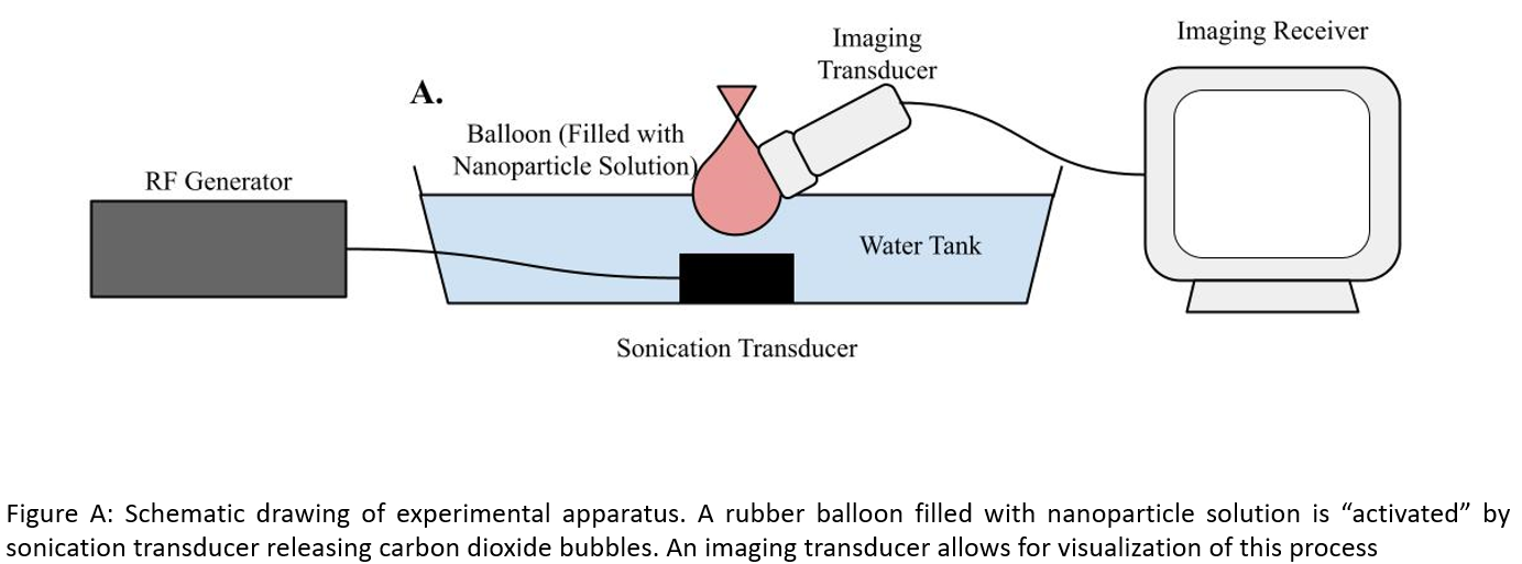
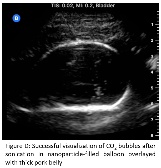
Methods: Polyethylenimine (PEI, 500-940 mg) was dissolved in solvent (DI water, ethylene glycol, or PBS; 10 mL), followed by addition of dry ice (50 g) as a CO2 source in a sealed Parr apparatus. Dry ice released CO2 and the reaction was stirred for 18 hours until it reached ambient temperature. A rubber balloon (simulating a bladder) was filled with either water only or the nanoparticle solution (Figure A). We used a prototype 1.1 MHz spherically focused, air-backed transducer (focal depth: 55 mm) or a prototype unfocused 1.66 MHz air-backed transducer, a computer-controlled RF generator (JJ&A Instruments) to provide bubble sonication, and a Butterfly iQ imaging system to simultaneously visualize the bubbles produced. We used an electrical power of 10 W with 10% duty cycle (1 ms on, 9 ms off) for a period of 5 seconds. Following this, a 37mm layer of pork belly was interposed between the ultrasound transducers and the experiment repeated.
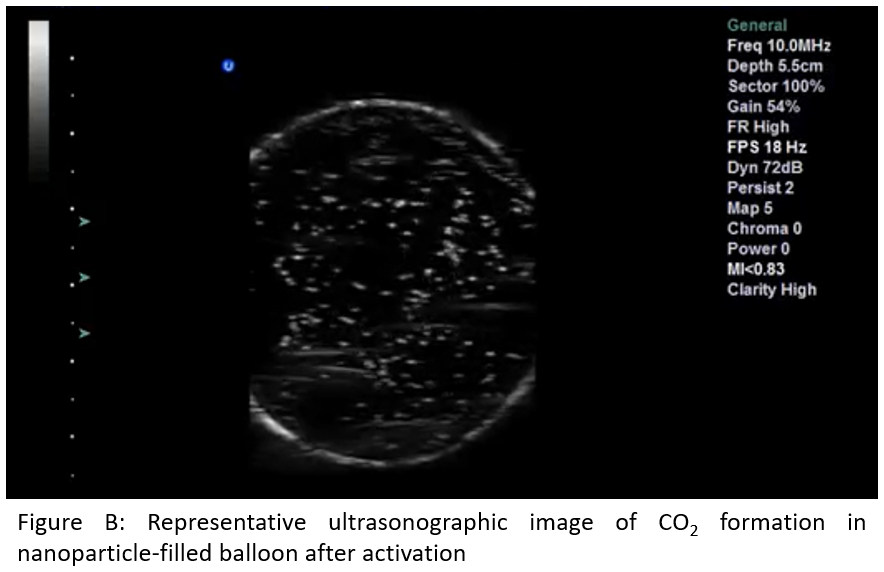
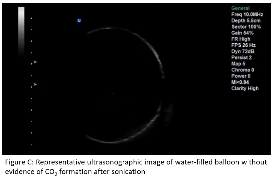
Results: Figures B and C show the nanoparticle and water solutions upon stimulation with ultrasound, respectively in the balloon-only model. Our work demonstrates the potential to produce and visualize microbubbles when applied to CO2-loaded nanoparticles. Additionally, visualization was successful with a 37 mm thick layer of pork belly interposed between the ultrasound and balloon (Figure D).
Conclusions: We have demonstrated the ability to selectively release covalently bound CO2 nanoparticles and utilize the CO2 bubbles as an ultrasound contrast agent. This is a major technical hurdle we have overcome in our quest to develop a catheter-free, radiation-free VCUG. Our next steps are to refine our nanoparticles, optimize ultrasound activation parameters, and test this modality in an animal model to detect VUR.
Back to 2023 Abstracts