
Identifying Urinary Tract Dilation Classification in Renal Bladder Ultrasound Image Utilizing Computer Vision Machine-Learning Algorithms for Neonatal Hydronephrosis
Hsin-Hsiao Scott Wang, MD, MPH, MBAn, Michael Li, MBAn, PhD, Andrew Zhang, BA (candidate), Anudeep Mukkamala, MD, Carlos Estrada, MD, MBA.
Boston Children's Hospital, Boston, MA, USA.
Objectives: The urinary tract dilation (UTD) classification system provides objective assessment for hydronephrosis in young children using renal ultrasound (US) images. However, the variable reporting method and radiologist’s experience make it highly challenging to uniformly derive UTD classifications. We seek to utilize computer vision (CV) algorithms to identify UTD classifications and detailed components from US.
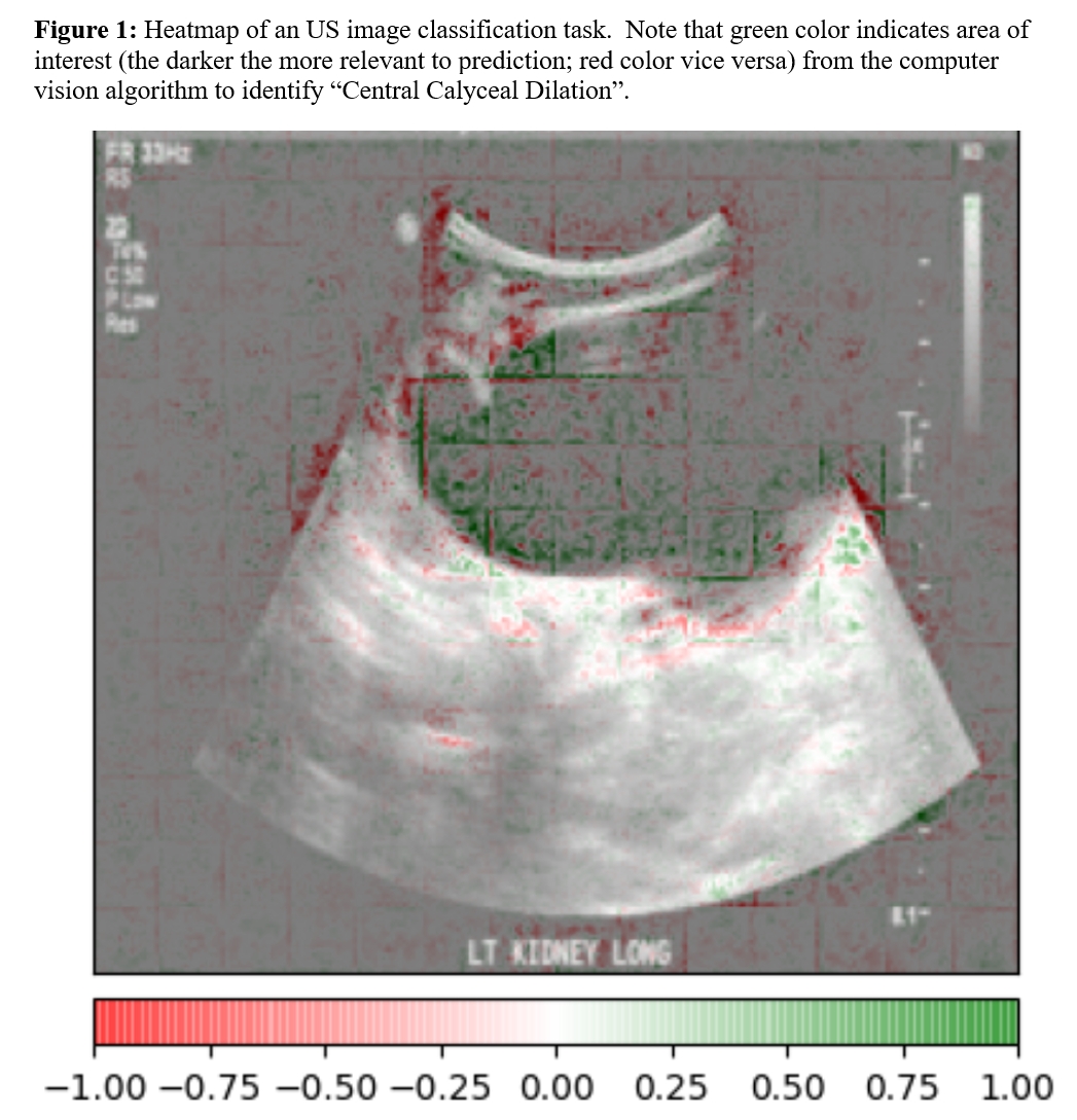
METHODS: US images from our institution were reviewed to identify infants aged 0-90 days undergoing early ultrasound for antenatal UTD during 2011-2022. The original US images, in Digital Imaging and Communications in Medicine (DICOM) format, were first preprocessed to convert to the standard JPEG format, and then reviewed by the research team physicians to create the ground truth of UTD components (including central calyceal dilation, peripheral calyceal dilation, abnormal ureter, abnormal parenchymal thickness, abnormal parenchymal appearance, and abnormal bladder as primary outcomes) used for P1/P2/P3 classifications. The JPEG Images were split into training/testing sets by an 80:20 ratio. Bidirectional Encoder Representations from Transformers (BERT) image representation models were used as the basis of the neural network classification model. The model was fine-tuned with a head consisting of a drop layer, a fully connected layer and a binary classification layer. The extracted UTD components were then further assembled to produce the standardized P1/P2/P3 classifications based on the UTD classification system definition. The model performance was then evaluated with out-of-sample testing image set. We further used explainable AI tool Captum to plot heatmaps to show the areas that are important for the model’s classification decision. Figure 1 shows the heatmap of an US image that is classified as central dilation by the model.
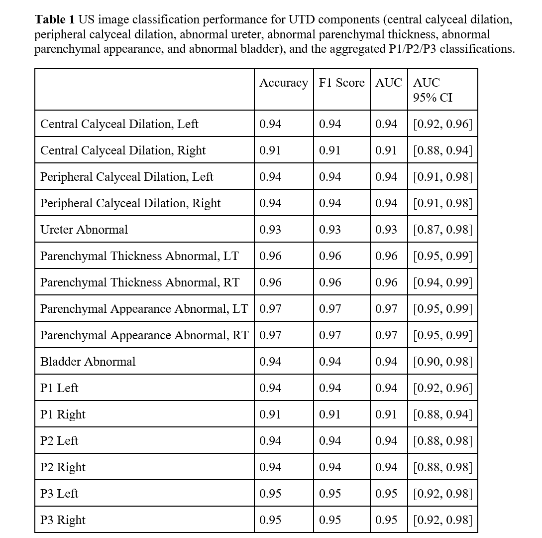
RESULTS: In total, over 48,000 US images (1104 US sessions, 1094 unique young [0-90 days] infants) were included in the study. The model performance for the out-of-sample testing image set is very high across UTD components and grades (AUC>0.91 for all). Accuracy, F1 score and AUC for UTD features and the P1/P2/P3 classifications in the out-of-sample testing image set are listed in Table 1.
CONCLUSIONS: By applying deep state-of-the-art CV neural networks, we developed a high-performing and scalable solution to identify UTD components and grades from US images. The CV model is explainable with heatmaps showing the areas on the images that are important for its decisions. This will allow future production of consistent UTD classification for hydronephrosis for clinical and research use.
Back to 2023 Abstracts